Introduction
Proximal tibia fractures, including tibial plateau fractures, are complex intra-articular injuries that involve the weight-bearing surface of the knee joint. These fractures account for approximately 1% of all adult fractures and constitute a significant challenge due to their complex anatomy and the biomechanical forces acting upon the knee. Epidemiologically, these fractures occur predominantly in two populations: younger males following high-energy trauma (e.g., road traffic accidents, sports injuries) and older females with osteoporotic bone who sustain low-energy injuries such as falls.
Treatment options range from conservative management for non-displaced fractures to surgical intervention involving open reduction and internal fixation (ORIF) with plates, screws, or less invasive stabilization systems (LISS). Surgical goals focus on anatomic reduction of the articular surface, restoration of mechanical alignment, and stable fixation to allow early mobilization.
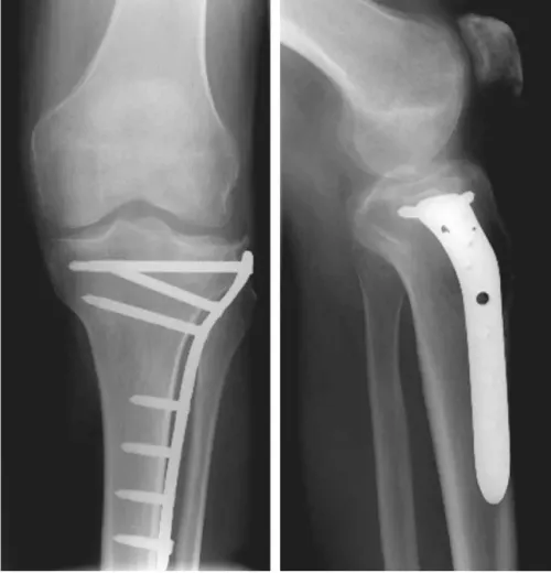
Proximal tibia Fracture fixation using Less Invasive Stabilization System (LISS)
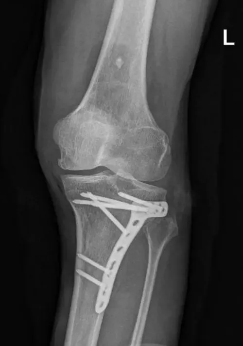
Proximal tibia Fracture fixed with T-shaped plate and screws.
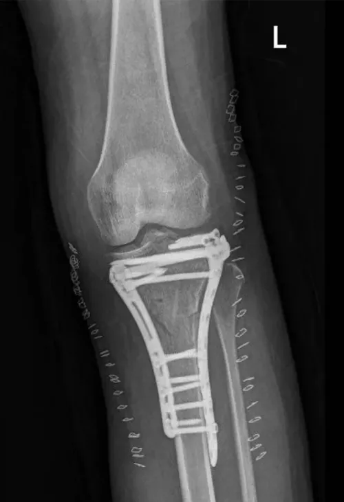
Proximal tibia Fracture fixed with dual plates and screws.
Despite advances in surgical fixation techniques, successful functional recovery heavily depends on an appropriate rehabilitation program. Early mobilization and range of motion (ROM) exercises have been advocated to prevent joint stiffness and promote cartilage nutrition. Weight-bearing protocols have historically been conservative (non-weight bearing or partial weight bearing for 6-12 weeks), but emerging evidence supports earlier weight bearing in selected cases without increased risk of fixation failure or loss of reduction. Rehabilitation protocols need to be individualized based on fracture type, fixation stability, and patient factors such as osteoporosis.
Phase 1: Immediate Postoperative Phase (Week 0-2)
- Goals:
- Protect surgical fixation and promote fracture healing.
- Control pain and inflammation.
- Prevent joint stiffness and muscle atrophy.
- Maintain cardiovascular fitness as tolerated.
- Interventions:
- Knee immobilization with brace in extension during ambulation
- ROM allowed: typically 0-90° flexion restriction depending on fracture/ligament injury).
- Cryotherapy and limb elevation (15-20 cm) to reduce swelling and pain.
- Neuromuscular electrical stimulation (NMES) of quadriceps to minimize muscle atrophy without stressing fixation.
- Non-weight bearing or toe-touch weight bearing (approx. 10 kg), depending on surgeon’s recommendation and fixation stability.
- Precautions:
- Avoid active knee flexion beyond surgeon’s prescribed range.
- Limit weight-bearing according to fixation stability.
- Monitor for signs of infection, fixation failure, or compartment syndrome.
- Exercises:
- Isometric quadriceps and hamstring contractions Gluteal sets
Hip abductor isometrics
-
- Ankle pumps and toe flexion-extension exercises to reduce venous thromboembolism risk Passive knee ROM within allowed limits.
Phase 2: Early Rehabilitation Phase (Weeks 3-6)
- Goals:
- Gradually increase knee ROM.
- Begin partial weight bearing as tolerated.
- Improve muscle strength and neuromuscular control.
- Maintain cardiovascular fitness.
- Interventions:
- Progress knee flexion gradually aiming for at least 90-120° of ROM by week 6.
- Continuous passive motion (CPM) devices may be used, though evidence on impact is mixed
- Begin partial weight bearing, typically progressing from toe-touch (~10 kg) to 20-30 kg as tolerated.
- Continue NMES if needed.
- Introduce hip abductor strengthening to assist knee joint loading and gait mechanics.
- Soft tissue mobilization and gentle stretching.
- Use of knee brace may continue if ligamentous injury or instability present .
- Precautions:
- Avoid full weight bearing unless cleared by surgeon.
- Avoid deep knee flexion or high impact activities.
- Monitor for pain and swelling.
- Exercises:
- Side-lying hip abduction.
- Straight leg raises
- Stationary cycling without resistance as tolerated
Phase 3: Progressive Rehabilitation Phase (Weeks 7-12)
- Goals:
- Achieve full knee ROM (0-130° or greater).
- Progress to full weight bearing.
- Improve muscle strength and endurance.
- Restore normal gait pattern.
- Enhance balance and proprioception.
- Interventions:
- Advance weight bearing to full as tolerated; many patients achieve full weight bearing by 9-12 weeks.
- Initiate closed kinetic chain exercises (e.g., mini squats within pain-free range)
- Balance and proprioceptive training.
- Gait training focusing on normalizing step length and cadence.
- Low-level laser therapy
- Precautions:
- Avoid high-impact activities.
- Watch for swelling or increased pain indicating overloading.
- Continue to protect fixation if fracture healing not confirmed radiographically.
- Exercises:
- Partial squats (0-30°) progressing to full squats.
- Step-ups and step-downs.
- Stair climbing training
- Hip abduction and extension strengthening.
- Single-leg stance and balance exercises.
- Cycling and swimming.
Phase 4: Advanced Rehabilitation Phase (3-6 Months)
- Goals:
- Restore full function and strength.
- Return to vocational and recreational activities.
- Optimize neuromuscular control.
- Interventions:
- Advance to dynamic and plyometric exercises as tolerated.
- Running progression and sport-specific drills for athletes.
- Continued muscle strengthening focusing on quadriceps, hamstrings, and hip muscles.
- Gait and balance re-training.
- Address any residual deficits in ROM or strength.
- Precautions:
- Avoid activities that cause pain or swelling.
- Monitor for signs of joint instability or mechanical symptoms.
- Exercises:
- Lunges, hops, and agility drills.
- Resistance training with weights.
- Endurance training.
Phase 5: Return to Full Activity (6-12 Months)
- Goals:
- Full return to pre-injury levels of activity.
- Maintain joint health and function.
- Prevent post-traumatic osteoarthritis.
- Interventions:
- Maintain strength and conditioning program.
- Regular functional assessments.
- Patient education on joint protection strategies.
- Precautions:
- Continued avoidance of extreme joint loading if advised.
- Monitor for signs of degenerative changes.
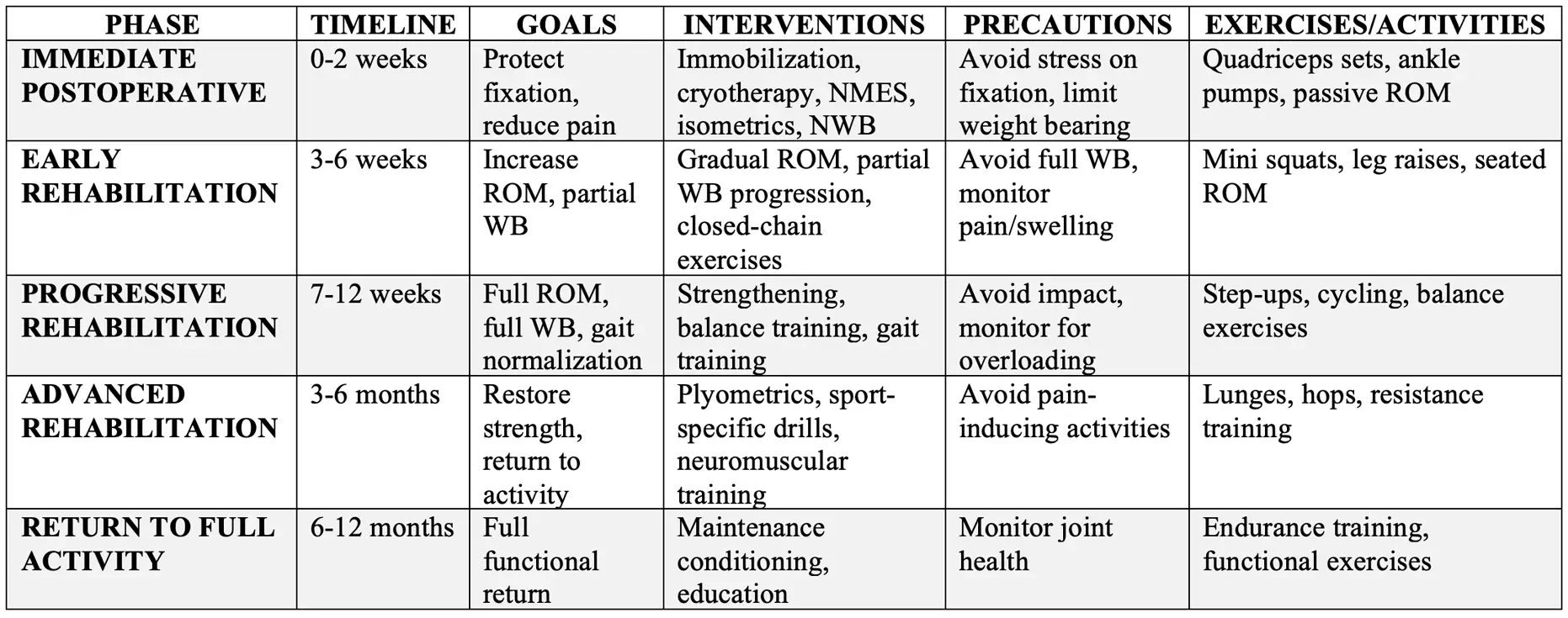
Summary Table of Rehabilitation Protocol
New Findings and Important Points
- Early Weight Bearing: Recent studies challenge traditional delayed weight-bearing protocols and suggest that early or immediate weight bearing (as tolerated) after stable fixation may be safe and beneficial, leading to faster functional recovery without increased complication rates.
- Patient Compliance: Surveys indicate variability in surgeons’ weight-bearing recommendations and patient adherence is often poor; rehabilitation plans should incorporate patient education and objective weight-bearing monitoring where possible.
- Muscle Strengthening: Quadriceps and hip abductor strengthening are essential for optimizing knee joint biomechanics during gait and reducing the risk of long-term osteoarthritis.
- Use of CPM: Continuous passive motion devices are commonly used but their impact on outcomes remains unclear.
- Nutritional Support: Supplementation with vitamin D, calcium, and joint-supportive nutraceuticals (glucosamine, chondroitin) may aid bone and cartilage healing.
Common Questions and Answers (For Patients)
1. How long will I need crutches after a proximal tibia fracture?
The length of time you need crutches depends on the severity and location of the fracture, whether you had surgery, the stability of the fixation, and how well the bone is healing.
Your surgeon will determine when you can begin putting weight on the injured leg, often based on follow-up X-rays and your clinical progress. Your physiotherapist can then help you safely progress from protected weight bearing to walking independently.
It is important not to increase weight bearing beyond your prescribed restrictions without clearance from your surgeon.
2. When can I put weight on my leg after a proximal tibia fracture?
Weight bearing is usually progressed gradually and depends on the type of fracture and how well the bone and surgical fixation are healing.
Some patients may initially be instructed to remain non-weight bearing or only partially weight bearing. As healing progresses and your surgeon confirms that the fracture is sufficiently stable, weight bearing can be increased.
Your rehabilitation should follow your surgeon’s weight-bearing instructions while progressively rebuilding strength and walking ability.
3. When will I be able to walk normally after a proximal tibia fracture?
Walking normally can take time because the knee and leg may remain weak and stiff even after you are allowed to fully bear weight.
Physiotherapy focuses on restoring knee movement, quadriceps strength, balance, coordination, and walking mechanics. As your strength and control improve, you will gradually progress from using an assistive device to independent walking.
The goal is to restore a safe and efficient walking pattern rather than simply stopping your crutches as quickly as possible.
4. How can I improve my knee movement after a proximal tibia fracture?
Knee stiffness is common following a proximal tibia fracture, particularly when the knee has been immobilized or weight bearing has been restricted.
Your rehabilitation program may include gentle range-of-motion exercises, quadriceps activation, and progressive strengthening. The amount and type of movement should be appropriate for your fracture and surgical restrictions.
Restoring knee extension and flexion is an important part of regaining normal walking and daily function.
5. When can I go up and down stairs normally?
Stairs require more strength and control than level walking and can remain challenging during the recovery period.
Initially, you may need to use a railing, crutches, or a modified stair pattern. As your knee movement, strength, balance, and weight-bearing tolerance improve, your physiotherapist will help you progress toward using stairs normally.
Your ability to manage stairs safely is more important than reaching a specific timeline.
6. When can I drive after a proximal tibia fracture?
You should return to driving only when you can safely control the vehicle and perform an emergency braking manoeuvre without significant pain or hesitation.
Your ability to drive depends on your knee movement, strength, reaction time, weight-bearing status, and which leg was injured. A brace or crutches may also affect your ability to drive safely.
Always follow your surgeon’s recommendations and check with your insurance provider before returning to driving.
7. When can I return to work after a proximal tibia fracture?
Your return-to-work timeline depends on your recovery and the physical demands of your job.
A desk-based job may allow an earlier return, while jobs involving prolonged standing, walking, stairs, kneeling, lifting, or heavy physical work may require a longer recovery period.
A gradual return to work with temporary modifications may be appropriate. Your physiotherapist can assess your functional abilities and help develop recommendations based on your specific job requirements.
8. When can I start exercising after a proximal tibia fracture?
Rehabilitation exercises usually begin during the early stages of recovery, but the type and intensity of exercise depend on your fracture and surgical restrictions.
Early exercises may focus on maintaining knee movement, activating the quadriceps, reducing stiffness, and maintaining strength in the rest of the leg. As the fracture heals and weight bearing increases, rehabilitation can progress to resistance training, balance exercises, functional strengthening, and cardiovascular exercise.
Your exercise program should gradually become more challenging as your bone, knee, and muscles become stronger.
9. When can I start squatting after a proximal tibia fracture?
Squatting places significant load through the knee and should be introduced gradually.
Initially, your rehabilitation may involve partial or supported squatting within a comfortable and appropriate range. As your fracture heals and your strength improves, you can gradually progress toward deeper squats and eventually loaded squatting when appropriate.
Your physiotherapist will consider your strength, knee movement, pain, swelling, and fracture healing before progressing this activity.
10. When can I start running after a proximal tibia fracture?
Running should not be started simply because a certain amount of time has passed.
Before returning to running, you should have adequate fracture healing, full or near-full knee movement, minimal swelling, sufficient strength, and good single-leg control.
Your physiotherapist can assess your readiness and develop a gradual return-to-running program. Higher-impact activities should only be introduced once your surgeon and rehabilitation team consider your bone and knee sufficiently prepared.
11. When can I return to sports after a proximal tibia fracture?
Returning to sports is a gradual process and depends on the severity of the fracture, your treatment, bone healing, strength, knee function, and the demands of your sport.
Low-impact activities such as cycling, swimming, walking, and certain fitness activities may be introduced earlier. Sports involving running, jumping, pivoting, or rapid changes in direction require a higher level of strength and control and usually take longer.
Before returning to full sport, you should demonstrate adequate strength, balance, movement control, and the ability to perform sport-specific activities without significant pain or swelling.
12. How long does rehabilitation take after a proximal tibia fracture?
Recovery varies significantly from person to person. Factors such as the fracture pattern, whether surgery was required, the stability of the fixation, bone healing, age, overall health, and physical demands of your daily activities can all affect recovery.
Some patients regain basic function within several months, while more complex fractures may require a longer rehabilitation period. Returning to higher-level activities and sports can take considerably longer.
Rather than focusing only on a specific number of weeks or months, rehabilitation should be guided by your fracture healing, knee movement, strength, balance, walking ability, and functional progress.
Reference:
- Ali, A. M., Burton, M., Hashmi, M., et al. (2003). Outcome of complex fractures of the tibial plateau treated with a beam-loading ring fixation system. Journal of Bone and Joint Surgery (British Volume), 85(5), 691–699.
- Arnold, J. B., Tu, C. G., Phan, T. M., et al. (2017). Characteristics of postoperative weight bearing and management protocols for tibial plateau fractures: Findings from a scoping review. Injury, 48(12), 2634–2642.
- Canton, G., Sborgia, A., Dussi, M., Rasio, N., & Murena, L. (2022). Early weight bearing in tibial plateau fractures treated with ORIF: A systematic review of literature. Journal of Orthopaedic Surgery and Research, 17(1), 261.
- Elsenosy, A. M., Yousef, A. S., Hassan, E., et al. (2025). Early Versus Delayed Weight-Bearing Following Tibial Plateau Fracture Surgery: A Systematic Review and Meta-Analysis. Cureus, 17(10), e95185.
- Gosling, T., Schandelmaier, P., Müller, M., et al. (2005). Single lateral locked screw plating of bicondylar tibial plateau fractures. Clinical Orthopaedics and Related Research, 439, 207–214.
- Granlund, A. S., & von Keudell, A. G. (2025). Weight bearing after surgical treatment of tibial plateau fractures – an international survey of orthopaedic trauma surgeons. Injury, 56, 112599.
- Hakukawa, S., Kashiwagura, K., Kishimoto, H., et al. (2025). Impact of quadriceps and hip abductor strengthening on knee joint biomechanics during gait after Schatzker type III tibial plateau fracture: A case study. Cureus, 17(1), e78092.
- Iliopoulos, E., & Galanis, N. (2020). Physiotherapy after tibial plateau fracture fixation: A systematic review of the literature. SAGE Open Medicine, 8, 2050312120965316.
- Ibrahim, M. A., Moustafa, M. M. A., Brismée, J. M., et al. (2025). Immediate weight-bearing after tibial plateau fractures internal fixation results in better clinical outcomes with similar radiological outcomes: A randomized clinical trial. International Orthopaedics, 49, 1245–1253.
- Kadam, A., Wani, S., Gadekar, G., et al. (2024). Effect of early weight bearing program with conventional physiotherapy on functional outcomes in surgically treated proximal tibia fracture: A pilot randomized controlled trial. Archives of Orthopaedic and Trauma Surgery, 144, 2481–2489.
- Phan, T. M., Arnold, J., Solomon, L. B., et al. (2017). Characteristics of postoperative weight bearing and management protocols for tibial plateau fractures: Findings from a scoping review. Injury, 48(12), 2634–2642.
- Smith, T. O., Hedges, C., & Schankat, K. (2010). A systematic review of the rehabilitation of LISS plate fixation of proximal tibial fractures. Advances in Physiotherapy, 12(2), 100–110.
- Zhu, Z., Wang, H., & Kang, R. (2025). Impact of systematic postoperative rehabilitation training on prognosis in patients with osteoporosis and tibial plateau fractures: A retrospective study. Current Problems in Surgery, 64, 101729.