Comprehensive Rehabilitation Protocol Following Ankle Lateral Collateral Ligament Reconstruction

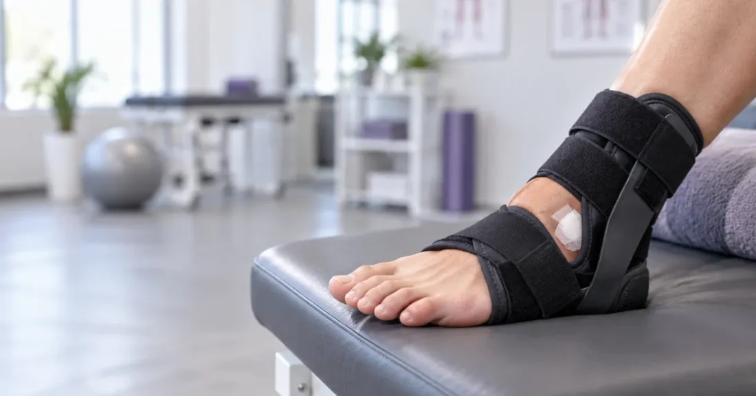
Patient's foot resting on a physical therapy bench wearing a supportive post-operative ankle brace.

Introduction

Ankle sprains are among the most common musculoskeletal injuries, representing up to 50% of lower extremity injuries in athletic populations and accounting for nearly 30% of all sports injuries in the United States. The lateral collateral ligament complex of the ankle, composed of the anterior talofibular ligament (ATFL), calcaneofibular ligament (CFL), and posterior talofibular ligament (PTFL), is particularly vulnerable due to anatomic and biomechanical predispositions. Injuries to the ATFL are most frequent because it has the lowest ultimate load among the lateral ligaments.

While most acute ankle sprains heal with conservative management, approximately 20% of patients develop chronic lateral ankle instability (CLAI), characterized by recurrent sprains, “giving way,” pain, swelling, and functional limitations. CLAI can be subdivided into mechanical instability (ligament laxity confirmed by physical and imaging exams) and functional instability (neuromuscular deficits without mechanical laxity).

Surgical intervention is indicated when nonoperative treatment fails after 3 to 6 months, especially in cases of mechanical instability. The modified Broström procedure and its variants are considered the gold standard for anatomic repair of lateral ankle ligaments due to their durability and preservation of normal joint kinematics. In cases with insufficient ligament tissue or high-demand athletes, anatomic reconstruction using tendon grafts (autograft or allograft) is preferred. Arthroscopic-assisted techniques have gained popularity but demonstrate similar outcomes to open procedures.

Importance of Rehabilitation

Postoperative rehabilitation is critical to restore ankle stability, strength, proprioception, and function, enabling patients to return to pre-injury activity levels safely. Rehabilitation balances protecting the surgical repair with avoiding joint stiffness, muscle atrophy, and delayed return to sport. Early controlled range of motion (ROM) and weight-bearing have been shown to improve functional outcomes and expedite return to activity without compromising stability or increasing complication rates. However, rehabilitation protocols vary widely, and standardization remains a need.


Rehabilitation Protocol

This protocol synthesizes evidence from multiple high-level studies and clinical guidelines.

Phase 1: Immediate Postoperative (Weeks 0–2)

Goals:

  • Protect surgical repair to allow initial ligament healing.
  • Control pain and swelling.
  • Prevent complications related to immobilization.
  • Maintain non-affected joint mobility.

Interventions:

  • Immobilization with a posterior splint or short leg cast/boot.
  • Weight-bearing: Immediate weight-bearing as tolerated (WBAT) in a boot is supported with suture-tape augmentation; otherwise, non-weight-bearing or partial weight-bearing protocols are acceptable depending on surgeon’s preference and type of reconstruction.
  • Elevation and cryotherapy for edema control.
  • Begin movement exercises for toes, knee, and hip to prevent stiffness.
  • Ankle dorsiflexion and plantarflexion range of motion may begin as early as postoperative day 2 when safe, especially with augmented repairs.

Precautions:

  • Avoid inversion and excessive ankle movements that stress the repair.
  • Prevent premature weight-bearing if advised by surgeon.

Exercise examples:

  • Toe curls and wiggles.
  • Isometric contractions of the quadriceps and hamstrings.
  • Gentle active ROM of toes, knee, and hip.
  • Ankle pumps (dorsiflexion/plantarflexion) within pain-free range.
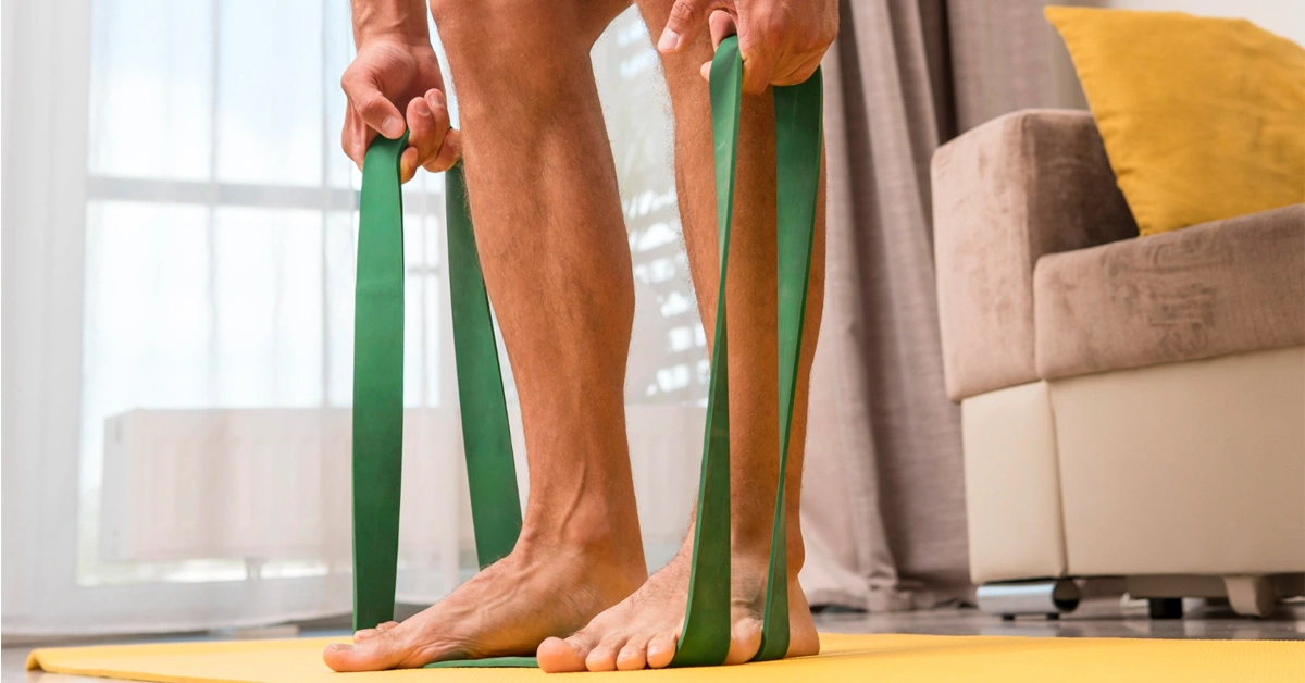
  • Non-weight-bearing isometric ankle eversion and inversion against resistance bands (without stressing repair).

Phase 2: Early Rehabilitation (Weeks 2–6)

Goals:

  • Gradual restoration of ankle ROM.
  • Progress weight-bearing safely.
  • Begin strengthening exercises focusing on peroneal muscles.
  • Initiate proprioceptive and balance training.
  • Maintain cardiovascular fitness with low-impact activities.

Interventions:

  • Transition from cast/splint to removable boot or ankle brace.
  • Progress from partial to full weight-bearing as tolerated (often by week 4–6).
  • Initiate physical therapy focused on controlled ankle ROM, peroneal strengthening, and proprioception.
  • Cardiovascular exercises such as stationary cycling and water therapy.

Precautions:

  • Avoid inversion stretching until after 3 months post-op to protect repair integrity.
  • Monitor for pain or swelling during increased activity.

Exercise examples:

  • Active ankle ROM exercises including dorsiflexion, plantarflexion, and eversion within a controlled range (inversion avoided or limited).
  • Isometric and isotonic peroneal muscle strengthening (e.g., resisted eversion with elastic bands).
  • Foot intrinsic muscle exercises (e.g., “short-foot” exercises).
  • Balance exercises progressing from double-leg stance to single-leg stance on stable surfaces.
  • Gait training to restore symmetrical walking.

Phase 3: Intermediate Rehabilitation (Weeks 6–12)

Goals:

  • Restore full ankle ROM.
  • Improve muscular strength and endurance.
  • Enhance dynamic balance and proprioception.
  • Begin low-impact functional activities.
  • Prepare for gradual return to running.

Interventions:

  • Discontinue boot; transition to ankle brace during ambulation.
  • Progressive strengthening with resistance exercises.
  • Advanced proprioceptive training on unstable surfaces.
  • Begin closed kinetic chain exercises (e.g., squats, lunges).
  • Initiate low-speed jogging and elliptical training when full weight-bearing without pain is achieved.

Precautions:

  • Avoid high-impact or agility drills until adequate strength and balance are demonstrated.
  • Ensure no swelling or pain with progression of activity.

Exercise examples:

  • Full active and passive ankle ROM in all planes.
  • Resistance band exercises for dorsiflexion, plantarflexion, inversion, and eversion.
  • Single-leg balance exercises on foam or wobble boards.
  • Heel raises progressing from double-leg to single-leg.
  • Closed kinetic chain strengthening: squats, lunges.
  • Low-impact cardio: cycling, elliptical.
  • Gradual introduction of jogging and treadmill walking.

Phase 4: Advanced Rehabilitation and Return to Sport (Weeks 12–16+)

Goals:

  • Achieve ≥90% strength and proprioception compared to contralateral limb.
  • Restore neuromuscular control for sports-specific activities.
  • Progress to high-impact and agility exercises.
  • Safe return to full sport participation.

Interventions:

  • Wean off ankle brace during sport-specific activities.
  • Plyometric training: bilateral progressing to unilateral hops.
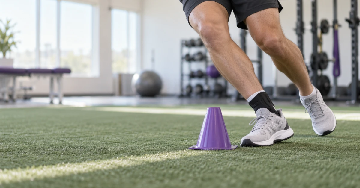
  • Agility drills: zigzag running, cutting, figure-of-8 runs.
  • Functional tests to assess readiness for return to sport (single-leg hop, Star Excursion Balance Test).
  • Continued strengthening and proprioceptive exercises.

Precautions:

  • Avoid premature return to competitive play without passing functional criteria.
  • Monitor for pain, swelling, or instability signs during advanced activities.

Exercise examples:

  • Plyometric drills: jump squats, hopping drills.
  • Agility drills: shuttle runs, lateral movements.
  • Sport-specific drills tailored to athlete’s demands.
  • Functional performance tests for strength and balance.
  • Dynamic balance training with perturbations.

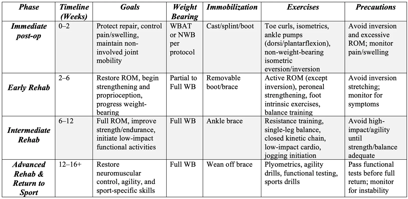
Summary Table of Rehabilitation Protocol

Rehabilitation protocol chart for lateral ankle ligament reconstruction showing recovery phases and exercises.

New Findings and Important Points

  • Early Weight Bearing: Studies, particularly those involving suture-tape augmented Broström procedures, support immediate or early weight-bearing postoperatively without compromising repair integrity, leading to earlier return to activity.
  • Early Range of Motion: Initiating controlled ankle dorsiflexion and plantarflexion motions as early as 2 days post-op improves outcomes and reduces joint stiffness.
  • No Increase in Complications: Early mobilization protocols do not appear to increase complication rates such as wound issues or repair failure.
  • Functional Testing: Use of objective functional tests (single-leg hop, heel raise, Star Excursion Balance Test) is essential in guiding safe return to sport.
  • Variability and Need for Standardization: There is significant variability in rehabilitation protocols, highlighting the need for standardized, evidence-based guidelines.

This protocol aims to provide clinicians and patients with a comprehensive, evidence-based guide for rehabilitation following ankle lateral collateral ligament reconstruction, incorporating the latest findings on early mobilization and functional recovery. Adherence to this staged progression, with attention to individual patient response, will optimize outcomes and facilitate a safe return to activity and sport.


Frequently Asked Questions

1. How soon can I start putting weight on my foot after ankle ligament surgery?

Depending on the surgical technique, especially if a suture-tape augmentation was used, you may be allowed to start putting weight on your foot immediately or within the first two weeks. Always follow your surgeon’s instructions.

2. When will I regain full range of motion in my ankle?

Controlled ankle movements typically begin within the first two weeks post-surgery, with full range of motion expected around 6 to 12 weeks, depending on individual healing and rehabilitation progression.

3. What exercises will help me recover after surgery?

Early exercises include toe curls, ankle pumps, and gentle isometric contractions. Later phases include strengthening with resistance bands, balance training, closed kinetic chain exercises, and sport-specific agility drills.

4. When can I return to sports or high-impact activities?

Return to sport usually occurs around 12 to 16 weeks post-surgery, once you have regained sufficient strength, balance, and pain-free range of motion, and have passed functional tests.

5. Are there risks with starting rehabilitation too early?

While early rehabilitation has many benefits, it is important to avoid excessive inversion or stress on the healing ligaments. Proper supervision and adherence to a structured protocol minimize risks.

6. How important is balance training in my recovery?

Balance and proprioceptive training are critical as they help restore neuromuscular control, reduce the risk of re-injury, and improve functional stability.


References

  1. Al-Janabi, M. M., Apostolides, M., Southgate, C., & Dhinsa, B. S. (2023). Early mobilization following elective ankle lateral collateral ligament reconstruction in adults. The Foot, 55, 101988. https://doi.org/10.1016/j.foot.2023.101988 [[1], [2]]
  2. Camacho, L. D., Roward, Z. T., Deng, Y., & Latt, L. D. (2019). Surgical management of lateral ankle instability in athletes. Journal of Athletic Training, 54(6), 639–649. https://doi.org/10.4085/1062-6050-348-18 [[19]]
  3. Clements, A., Belilos, E., Keeling, L., Kelly, M., & Casscells, N. (2021). Postoperative rehabilitation of chronic lateral ankle instability: A systematic review. Sports Medicine and Arthroscopy Review, 29(2), 146–152. https://doi.org/10.1097/jsa.0000000000000326 [[30]]
  4. Hermanns, C., Coda, R., Cheema, S., Vopat, M. L., Bechtold, M., Tarakemeh, A., … & Vopat, B. G. (2020). Review of variability in rehabilitation protocols after lateral ankle ligament surgery. Kansas Journal of Medicine, 13, 152–159. https://doi.org/10.17161/kjm.vol13.13838 [[38]]
  5. Martin, K. D., Andres, N. N., & Robinson, W. H. (2021). Suture tape augmented Broström procedure and early accelerated rehabilitation. Foot & Ankle International, 42(2), 145–150. https://doi.org/10.1177/1071100720959015 [[46]]
  6. Miyamoto, W., Takao, M., Yamada, K., & Matsushita, T. (2014). Accelerated versus traditional rehabilitation after anterior talofibular ligament reconstruction for chronic lateral instability of the ankle in athletes. American Journal of Sports Medicine, 42(6), 1441–1447. https://doi.org/10.1177/0363546514527418 [[52]]
  7. Pearce, C. J., Tourné, Y., Zellers, J., Terrier, R., Toschi, P., & Grävare Silbernagel, K. (2016). Rehabilitation after anatomical ankle ligament repair or reconstruction. Knee Surgery, Sports Traumatology, Arthroscopy, 24(4), 1130–1139. https://doi.org/10.1007/s00167-016-4051-z [[59]]
  8. Song, Y., Li, H., Sun, C., Zhang, J., Gui, J., Guo, Q., … & Chen, S. (2019). Clinical guidelines for the surgical management of chronic lateral ankle instability: A consensus reached by systematic review of the available data. Orthopaedic Journal of Sports Medicine, 7(9), 2325967119873852. https://doi.org/10.1177/2325967119873852 [[69]]
  9. Vopat, M. L., Wendling, A., Lee, B., Hassan, M., Morris, B., Tarakemeh, A., … & Vopat, B. G. (2021). Early versus delayed mobilization post-operative protocols for primary lateral ankle ligament reconstruction: A systematic review and meta-analysis. Kansas Journal of Medicine, 14(1), 141–148. https://doi.org/10.17161/kjm.vol1415028 [[81]]