

Introduction
Distal Radius Fracture Overview
Distal radius fractures (DRFs) are among the most common fractures encountered in orthopedic practice, especially prevalent in adults over 50 years and exhibiting a bimodal distribution affecting elderly populations due to low-energy falls and younger individuals from high-energy trauma. The distal radius is a critical structural component of the wrist, characterized by a rectangular cross-section of cancellous bone prone to fractures, particularly at its metaphyseal region. Post-fracture complications can include wrist dysfunction, stiffness, pain, and secondary conditions such as complex regional pain syndrome (CRPS) and carpal tunnel syndrome (CTS).
Treatment Options
Treatment ranges from nonoperative management with casting or splinting to surgical intervention, including open reduction and internal fixation (ORIF) with volar locking plates, external fixation, or percutaneous pinning. Volar locking plate fixation has become increasingly favored for unstable or displaced fractures due to its biomechanical stability and facilitation of early mobilization. However, there remains debate regarding immobilization duration and the timing and nature of postoperative rehabilitation.
Importance of Rehabilitation
Rehabilitation after DRF is essential to restore wrist mobility, strength, and function, reduce pain and edema, and prevent complications such as stiffness and CRPS. Early mobilization and structured therapy have shown to enhance short-term outcomes in range of motion (ROM), grip strength, and patient-reported function. However, evidence regarding the superiority of supervised physiotherapy over home exercise programs (HEP) remains inconclusive, with patient-specific factors influencing the optimal rehabilitation approach. Rehabilitation protocols should be individualized, considering fracture type, treatment modality, patient age, and comorbidities.
Rehabilitation Protocol
Phase One: Early Motion Phase (0–4 weeks post-injury/surgery)
Goals:
- Educate patient on fracture rehabilitation and recovery expectations.
- Protect fracture site with immobilization.
- Minimize pain and edema.
- Maintain ROM of adjacent joints (shoulder, elbow, fingers).
- Prevent wrist, finger stiffness, and complications like CRPS.
- Maintain scapular stability and overall aerobic capacity.
Interventions:
- Immobilization using plaster cast or removable splint (typically 0-2 weeks post-surgery; longer for conservative treatment).
- Elevation of the limb above heart level to reduce swelling.
- Cryotherapy for pain and edema control .
- Active ROM (AROM) exercises for shoulder, elbow, cervical spine, and fingers.
- Pain-free passive ROM (PROM) for wrist and hand starting ~2 weeks if stable.
- Soft tissue mobilization for proximal muscle relaxation.
- Submaximal isometric contractions of wrist flexors/extensors starting after 2 weeks.
- Breathing and aerobic exercises.
- Edema control techniques: compression gloves, manual lymphatic drainage (MLD), massage, kinesiotaping.
Precautions:
- Avoid active wrist movements if fracture stability is not confirmed.
- Monitor for signs of CRPS or nerve involvement.
- Follow surgeon’s guidelines on immobilization duration and weight-bearing restrictions.
Exercise Examples:
- Finger flexion/extension and opposition exercises (e.g., intrinsic plus position, fist closure).
- Shoulder and elbow AROM exercises.
- Isometric wrist flexor/extensor contractions.
- Scapular mobilization and stabilization exercises.
- Edema management: elevation, compression gloves, gentle massage.
Phase Two: Active Motion Phase (5–8 weeks post-injury/surgery)
Goals:
- Maintain and improve muscle properties and wrist/hand mobility.
- Reduce edema and manage pain.
- Improve joint play and neuromuscular control.
- Enhance strength and endurance of wrist and forearm muscles.
- Restore scapulohumeral rhythm and proprioception.
Interventions:
- Removal or reduction of immobilization devices during exercises.
- Hot water fomentation (if swelling subsided).
- Continued edema management (compression gloves, edema mobilization).
- Desensitization exercises (varying textures, temperatures).
- Joint mobilizations (Maitland grades I and II, Mulligan mobilizations with movement).
- Introduce active-assisted ROM and active ROM of wrist and hand.
- Start mild resistance exercises shoulder & elbow.
- Proprioceptive training (vestibular ball exercises, quadruped weight-bearing).
- Functional exercises: towel crumpling, finger walking, tendon gliding, tenodesis exercises.
Precautions:
- Progress resistance exercises cautiously to avoid fracture site compromise.
- Monitor pain levels to avoid exacerbation.
- Avoid high-impact or load-bearing activities until adequately healed.
Exercise Examples:
- Active wrist flexion/extension, radial/ulnar deviation, supination/pronation.
- Proprioceptive exercises (e.g., vestibular ball, balance training with BOSU ball).
- Resistance training with exercise bands, light weights.
- Tendon gliding and differential tendon stretching.
- Functional hand tasks (pegboard activities, gripping, pinching).
Phase Three: Return to Activity Phase (9–12 weeks post-injury/surgery)
Goals:
- Achieve full, pain-free ROM at wrist, elbow, and shoulder.
- Increase muscle strength and endurance.
- Improve proprioception, neuromuscular control, and dynamic stability.
- Facilitate return to daily functional activities and work/sports.
Interventions:
- Progressive resistance strengthening (weights, exercise bands, isokinetic dynamometry).
- Functional training simulating activities of daily living (ADLs).
- Plyometric and power training (e.g., medicine ball throws, battle ropes).
- Aquatic therapy for strength and endurance.
- Continued joint mobilizations (grades III and IV).
- Advanced proprioceptive and balance training.
- Blood flow restriction (BFR) therapy as adjunct for strength gains, especially in older adults.
Precautions:
- Avoid overloading the wrist in presence of malunion or instability.
- Ensure adequate healing before high-impact exercises.
- Monitor for signs of recurrent pain or swelling.
Exercise Examples:
- Wrist and forearm curls with progressive overload.
- Plyometric drills (punching, medicine ball throws).
- Functional gripping and fine motor coordination exercises.
- Aquatic therapy exercises for strength and mobility.
- Isokinetic strengthening and BFR training protocols.
Additional Important Points
- Early Mobilization: Initiating wrist motion within 3-5 days post-surgery and starting strengthening exercises as early as 2 weeks postoperatively can lead to earlier return to function without increasing complications.
- Weight Bearing: Early weight-bearing through volar plates is generally safe and does not compromise fracture healing. Grip forces should be limited initially (<37.5 lb) to avoid implant overload.
- Supervised Therapy vs Home Exercise: Supervised therapy combined with home exercises is more beneficial for older adults and those with complications, while younger and uncomplicated cases might do well with home exercise programs alone.
- Edema Management: Manual lymphatic drainage and compression gloves can provide short-term benefits in edema control and improve ROM and function.
- Sensorimotor Training: Incorporation of graded motor imagery and mirror therapy can improve pain, proprioception, and function during early rehabilitation.
- Manual Therapy: Mobilization with movement and Maitland oscillations can enhance ROM and reduce pain when applied appropriately.
- Therapeutic Modalities: Use of light therapy (e.g., low-level laser, PEMF), cryotherapy, and warm whirlpool supports pain relief and functional recovery, though evidence on mechanical modalities like CPM and IPC is conflicting.
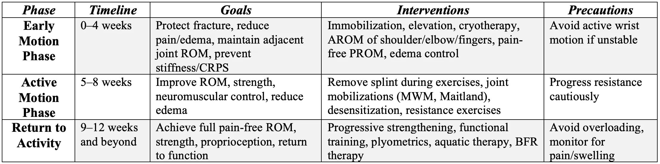
Summary Table of Rehabilitation Protocol
Frequently Asked Questions
1. How soon after a distal radius fracture can I start moving my wrist?
Early wrist motion can often begin within 3-5 days after surgery if your fracture is stabilized, but this depends on your surgeon’s advice and fracture stability.
2. Will starting exercises early increase my risk of complications?
Research shows that early rehabilitation does not increase complications or risk of losing fracture reduction when done under proper guidance.
3. Do I need supervised physiotherapy, or can I just do home exercises?
Supervised therapy is generally recommended for older adults or those with complications. Younger or uncomplicated cases can often achieve similar outcomes with a well-structured home exercise program.
4. How long will the rehabilitation process take?
Most patients regain functional wrist movement and strength within 3-6 months, although some exercises and strengthening may continue beyond this period.
5. What exercises should I do at home?
Early exercises include finger and shoulder ROM, progressing to wrist flexion/extension, and later strengthening with resistance bands and functional activities like gripping and pinching.
6. Can I put weight on my wrist early after surgery?
Yes, with stable fixation using volar locking plates, gradual weight-bearing and strengthening can start early (around 2 weeks), but heavy lifting should be avoided until cleared by your therapist and surgeon.
References
- Bruder, A. M., Taylor, N. F., Dodd, K. J., & Shields, N. (2013). Physiotherapy intervention practice patterns used in rehabilitation after distal radial fracture. Physiotherapy, 99(3), 233–240. https://doi.org/10.1016/j.physio.2012.09.003 [1]
- Brehmer, J. L., & Husband, J. B. (2014). Accelerated rehabilitation compared with a standard protocol after distal radial fractures treated with volar open reduction and internal fixation: a prospective, randomized, controlled study. The Journal of Bone and Joint Surgery. American Volume, 96(19), 1621–1630. https://doi.org/10.2106/JBJS.M.00860 [134]
- Zhou, Z., Li, X., Wu, X., & Wang, X. (2024). Impact of early rehabilitation therapy on functional outcomes in patients post distal radius fracture surgery: a systematic review and meta-analysis. BMC Musculoskeletal Disorders, 25(198). https://doi.org/10.1186/s12891-024-07317-0 [148]
- Deshpande, S. S., Ghodke, P. P., Kale, S. Y., Brahmbhatt, H. M., Shah, H. M., & Deshpande, S. (2026). Enhancing functional outcomes in distal end radius fracture rehabilitation. Journal of Orthopaedic Case Reports, 16(1), 278–287. https://doi.org/10.13107/jocr.2026.v16.i01.6680 [145]
- Mehta, S. P., Karagiannopoulos, C., Pepin, M.-E., Ballantyne, B. T., Michlovitz, S., MacDermid, J., & Grewal, R. (2024). Distal radius fracture rehabilitation: clinical practice guidelines linked to the International Classification of Functioning, Disability, and Health from the Academy of Orthopaedic Physical Therapy and Academy of Hand and Upper Extremity Physical Therapy of the American Physical Therapy Association. Journal of Orthopaedic & Sports Physical Therapy, 54(9), CPG1–CPG78. https://doi.org/10.2519/jospt.2024.0301 [136]
- Quadlbauer, S., Pezzei, C., Jurkowitsch, J., Rosenauer, R., Pichler, A., Schättin, S., Hausner, T., & Leixnering, M. (2020). Rehabilitation after distal radius fractures: is there a need for immobilization and physiotherapy? Archives of Orthopaedic and Trauma Surgery, 140(5), 651–663. https://doi.org/10.1007/s00402-020-03367-w [133]
- Ikpeze, T. C., Smith, H. C., Lee, D. J., & Elfar, J. C. (2016). Distal radius fracture outcomes and rehabilitation. Geriatric Orthopaedic Surgery & Rehabilitation, 7(4), 202–205. https://doi.org/10.1177/2151458516669202 [145]
- Handoll, H. H., & Elliott, J. (2015). Rehabilitation for distal radial fractures in adults. Cochrane Database of Systematic Reviews, (9), CD003324. https://doi.org/10.1002/14651858.CD003324.pub3 [101]